Chronic Lower Limb Ischaemia
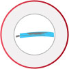
Peripheral Arterial Disease with narrowing or blockages in the arteries supplying the lower limbs causes chronic limb ischaemia.
Do I have Chronic Limb Ischaemia?
Some of the common symptoms are
- Intermittent Claudication
- Rest-pain
- Ulcers and Necrosis (black toes)
What is Intermittent Claudication?
Intermittent claudication is a condition characterised by pain and/or cramps in the lower leg muscles while walking and is relieved during rest. This type of pain that comes on with exertion and disappears with rest is referred to as intermittent claudication. When walking and exercising, the calf muscles need more oxygen to function, but the reduced blood flow does not carry enough oxygen. The pain may be felt as a dull ache or throbbing feeling.
When you present to our clinic with these symptoms, the diagnosis of claudication is made by a combination of clinical examination and a wide range of imaging tests (ultrasound, CT and MRA scans), and also by measuring and comparing blood pressures in the leg and arm (ankle-arm index).
To treat intermittent claudication, initially your doctor will ensure that you are medically optimised and the risk factors for PAD are addressed. This will typically include starting a blood thinner such as Aspirin and also a cholesterol lowering agent called statin. Statin is often prescribed as it has been shown by research to improve the function of the inner cell lining of the blood vessel wall. Although these medications will not improve the distance you can walk without claudication pain, it has been shown to reduce the incidence of heart attacks and strokes in the future. In-addition it is also important to stop smoking as there is a lot of evidence that quitting smoking reduces progression of PAD. Healthy diet, weight reduction and regular exercise will also help improve the longterm outcome from PAD.
Invasive treatment is not generally recommended or required for patients with claudication. It may be necessary only if you do not respond to conservative treatment and the claudication symptoms are severely interfering with your daily activities and quality of life. Angioplasty is a keyhole procedure (endovascular surgery) that opens up narrowed and blocked arteries by inflating a small balloon inserted via the groin. After an angioplasty, a stent (wire mesh tube) may be placed within the artery to hold the artery open. Your surgeon may perform a bypass surgery, where a graft (healthy blood vessel removed from another part of the body) is attached to re-route blood away from the block.
Rest Pain, Ulcers and Necrosis or (Critical Limb Ischaemia)
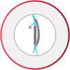
Rest pain is when the patient experiences pain in the legs especially the toes of the feet at all times. The pain is often worse at night time when trying to sleep with the legs up in the bed. These symptoms are a progression from intermittent claudication as the arterial disease advances and the blood supply to the legs is severely reduced. Often patients have to use very strong medications to get relief from pain symptoms. Minor knocks and bruises in the feet may struggle to heal due to the lack of adequate blood supply and nutrition. These then become ulcers and the tips of the toes may turn black (necrosis). Rest pain, ulcers and necrosis combined with reduced pressures (ABI) in the legs is referred to as Critical Limb Ischaemia(CLI). CLI is an indicator that the blood supply to the leg is severely depleted and here invasive intervention is warranted on an urgent basis to ensure that the leg does not deteriorate any further.
Common investigations for the diagnosis of Chronic limb ischaemia
ABI
What is Ankle-Brachial Index (ABI)?
The Ankle-Brachial Index (ABI) is a non-invasive diagnostic test performed to determine the severity of peripheral artery disease (PAD), a condition of narrowing or blocking of the arteries in your legs or arms. PAD also increases the risk of stroke and heart attacks. The ankle-brachial index is determined by comparing the blood pressures of the ankle and the arm. A low ABI value indicates narrowing of the peripheral arteries.
What are the indications?
The ABI test is indicated to determine the severity of PAD. It may also be recommended to check the efficacy of prescribed therapy (medicines, exercise, angioplasty or surgery) for PAD.
How is ABI performed?
Ankle-brachial index test is performed at the doctor’s office and doesn’t require any specific preparation. You will have to lie on the table; your doctor will use a standard blood pressure cuff to measure the pressure in your arms and also in the arteries at the ankle. A hand-held ultrasound device may be pressed on your skin to hear the pulse. The ankle-brachial index is calculated by dividing the highest blood pressure at the ankle by the highest blood pressure at the arms. The test is sometimes performed at rest and after exercise.
How are the results of ABI interpreted?
The normal value of ABI test at rest is 1.0 to 1.2 and it indicates absence of narrowed or blocked peripheral arteries. The following range of ABI values indicate the condition of arteries as below:
- ABI of 0.5 to 0.9 indicates narrowing or blocked arteries which can result in pain while exercising
- ABI of less than 0.4 indicates severe PAD or Critical Limb Ischaemia
- ABI of more than 1.2 indicates stiff arteries
If you are diabetic or have chronic kidney disease, the ABI test measured by standard procedure may not be accurate due to calcification (hardening) of the arteries. In such patients, measuring blood pressure of the toe (toe pressure) may provide more accurate results.
Duplex Ultrasound
What is Duplex Ultrasound?
Sonography or ultrasound is a diagnostic non-invasive test that creates images of the structures within the body with the help of reflected sound waves.
Duplex sonography is a process which combines regular ultrasound (image the structure of blood vessels) and Doppler ultrasound (images the flow of blood) to show abnormalities in blood vessels which affect blood flow.
Abdominal duplex sonography is performed to evaluate the important vessels (aorta and iliac arteries) passing through the abdomen. These arteries deliver blood to the major organs in the body. The test provides details such as
- blood flow in the arteries
- speed of the blood flow
- diameter of a blood vessel and degree of narrowing if present
The abdominal duplex ultrasound procedure is performed after overnight fasting so that bowel gases does not interrupt the ultrasound imaging. During the procedure, you will lie on a table and a gel is applied to the abdominal area to help the transmission of sound waves. A transducer is then moved over the abdomen to capture images of the blood vessels. Your surgeon then examines the images for abnormalities in blood vessels.
This procedure is a non-invasive, accurate and safe method of diagnosis and does not have any side effects.
CT Angiogram
What is CT Angiography?
Blood circulates in the human body through blood vessels. The deposition of calcium or plaques in these vessels can cause narrowing and blockages. These can be identified by CT angiography. This is a diagnostic test that involves the injection of a contrast dye into a superficial vein and this helps to visualise the blood flow within the lumen of the artery and vein.
Computerised tomographic angiography or CT angiography is a test that combines the technology of a CT scan (X-ray imaging that take images of the body in the form of slices) and angiography to create images of the blood vessels.
What are the indications and contraindications?
CT angiography is indicated to diagnose narrowing, blocks or bulging of blood vessels .
However, CT angiography is not recommended if you are allergic to iodine or shellfish. Kindly inform your physician regarding the same as it can increase the risk of allergies to the contrast dyes. Medications can be prescribed to reduce the risk of these allergies. CT angiography test is not recommended if you are pregnant as the rays may harm the foetus or if you have severe kidney disease as the dye can further damage the already sub-optimal function of the kidney.
Is there any specific preparation for this investigation?
Your physician might ask you to avoid foods and liquids up to 4 hours prior to the procedure. You may be allowed to continue medications that you are taking, but it is always advisable to confirm with your doctor prior to the examination.
Procedure
During CT angiography, a contrast dye is injected into your vein (mostly in the arm) with an injector machine which monitors the time and rate of the injection. This might make you feel warm or sick in your stomach.
You will be made to lie still on the scanning table in order to get good quality images. The scanning equipment consists of an X-ray tube which passes narrow beams of X-ray in the form of an arc over the body. The X-ray beams reflect on a detector present just opposite to the X-ray source. This machine can take about 1,000 pictures in a very short span of time and transmit them to a computer which converts it into 3D images. The position of the scanning table can be adjusted if images from different angles need to be taken. During the procedure, you may be asked to hold your breath for about 10 to 25 seconds as breathing may blur the image. The entire procedure may take about 20 minutes to 1 hour to complete.
Post-procedure care
After CT angiography, you will be advised to drink lots of fluids to flush out the contrast dye and avoid dehydration. There are no major restrictions and you will be able to resume your normal activities soon after the procedure.
Complications?
CT angiography may cause allergic reactions such as itching, difficulty in swallowing or breathing or flushing due to the contrast dye. These reactions should be reported to your doctor immediately. The contrast dye may also leak under the skin at the site of injection causing pain, swelling or redness. If you have any existing kidney problems, the contrast dye may cause further damage to the kidney function depending on the amount of dye used. Since CT angiography is an X-ray test, the radiation emitted might damage the body cells with repeated exposure.
However, under a skilled specialist, CT angiography can be a safe and successful investigation to diagnose diseases involving the arteries and veins.
MR Angiogram
What is Magnetic Resonance Angiography (MRA)?
This is a sophisticated non-invasive investigation that helps visualize the arterial and venous system. It relies on a large external magnetic field which magnetizes the charged particles within the body called protons. This in-turn can be manipulated and the vibrations from protons are tapped by sensitive receivers to process the final image of the arteries and veins. A special type of contrast or dye can also be used in MRI to visualize the arteries and veins without exposing the patient to iodine based dye. Modern MR scanners provide high quality images without exposing the patient to radiation. It has now become the imaging technique of choice for PAD in many centres.
Indications and contraindications
MRA is a sensitive investigation which is used to diagnose arterial abnormalities such as narrowing and blocks. It avoids the use of radiation and iodine based contrast agents or dye. Hence, it may be better suited than CT Angiography in some patients. However, it cannot be used in patients with Pacemaker or certain types of metallic implants like prosthetic heart valves, intracranial aneurysm clips or metallic foreign bodies in the eye. Patients on dialysis and kidney failure risk the complication of Nephrogenic Systemic Fibrosis (NSF) and MRA should be avoided in these patients.
Procedure
It is important to inform the clinician if you any metallic implant and you are required to fill in a questionnaire to ensure that you comply with the safety requirements for an MRI. You will lie down on a table that slides into a large magnet shaped tube. It is important to lie as still as possible during the examination to obtain a clear image. You may have plastic coils placed over you during this test. The staff can communicate with you during the test through a microphone in the room. The test may take between 30 and 60 minutes to complete and you may also receive a dye through a cannula in your arm. It is normal to expect loud bangs during the test which are noises made by the MRI machine.
What are the indications for treatment of Chronic Limb Ischaemia
- Rest pain with ulcerations and/ or necrosis (black) of toes
- Lifestyle limiting claudication